New Patients
Existing Patients
New Patients
Existing Patients
New Patients
Existing Patients
New Patients
Existing Patients
New Patients
Existing Patients
New Patients
Existing Patients
New Patients
Existing Patients
New Patients
Existing Patients

An intraoral camera is a compact, pen-sized imaging device designed to capture high-resolution, full-color images from inside the mouth. Unlike traditional external photos, these cameras are built to navigate tight spaces and present clear, magnified views of individual teeth, gum tissue, and other soft tissues. The result is a level of visual detail that helps clinicians spot surface irregularities, early wear, cracks, and areas of inflammation that may be hard to see with the naked eye alone.
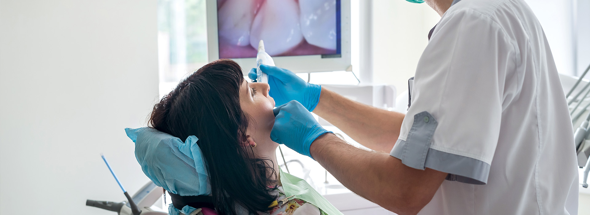
When used during an exam, the intraoral camera displays live video or still images on a chairside monitor, allowing both clinician and patient to examine the same view simultaneously. This real-time visualization helps the dental team evaluate the condition of restorations, detect spots of discoloration, and assess hard-to-reach interdental areas. The imagery is particularly useful for documenting subtle changes over time that may not be obvious during a cursory visual inspection.
Because the camera delivers crisp, color-accurate photos, it can reveal differences in texture and shading that assist with differential diagnosis. While it does not replace other diagnostic tools such as radiographs or CBCT when those are indicated, the intraoral camera adds a valuable optical layer to the diagnostic process, supporting more informed clinical decisions.
High-quality intraoral images serve as a practical extension of the oral exam, providing objective visual evidence to guide treatment planning. Clinicians use these images to map decay progression, evaluate margins of crowns and fillings, and identify cracked tooth patterns. Because the images can be paused, reviewed, and annotated, they become reference points during case discussions and when outlining treatment options.
The availability of detailed photos improves interdisciplinary care by making it easier to explain findings to dental specialists or laboratory technicians involved in restorations. For example, a clearly captured image of a fracture line or a failing margin helps a restorative dentist or lab technician design a targeted approach that preserves tooth structure and improves fit and function. In complex cases, intraoral imaging complements charting and radiographs to create a more complete clinical picture.
In practice, this translates to treatment plans that are more specific and better tailored to each patient’s needs. Images can illustrate why a conservative repair is advisable, when a restoration should be monitored, or when more extensive intervention is warranted. The visual evidence also supports accurate recordkeeping, which is important for continuity of care over time.
One of the most significant benefits of intraoral cameras is their value as a communication tool. When patients can see a magnified image of a problem area, abstract descriptions become concrete, and the rationale behind clinical recommendations becomes easier to grasp. This shared visual reference reduces uncertainty and empowers patients to participate more actively in decisions about their care.
The use of chairside images also helps demystify routine and restorative procedures. Instead of relying on verbal descriptions alone, clinicians can show the exact surfaces involved, point out areas of concern, and demonstrate how proposed treatments will address those issues. For many patients, seeing is the turning point from passive acceptance to informed consent based on clear evidence.
Moreover, intraoral photography is helpful for monitoring behavior-driven changes, such as enamel wear from grinding or localized gum recession from aggressive brushing. When patients observe the visual consequences of certain habits, they are often more motivated to adopt protective behaviors or follow preventive recommendations provided by the dental team.
Images captured with an intraoral camera are typically stored in the patient’s digital chart as part of the permanent record. These files create a visual timeline that clinicians can reference during follow-up visits, enabling side-by-side comparisons that reveal subtle progressions or improvements. Maintaining visual documentation supports objective clinical judgments and enhances the continuity of care when multiple providers are involved.
Because intraoral images are sharable in secure digital formats, they facilitate collaboration with specialists, dental laboratories, and other members of the care team. When sending a case for consultation or prosthetic fabrication, these photos provide additional context that complements written notes and radiographs. Clear imagery helps ensure that everyone involved has the same understanding of anatomy, margins, and areas requiring attention.
Secure storage and transfer protocols protect patient privacy while making exchange efficient. The production-quality images that intraoral cameras provide also reduce miscommunication, which can shorten turnaround times for lab work and lead to restorations that better match clinical expectations.
Intraoral cameras are noninvasive tools and are used under standard infection-control protocols similar to other intraoral instruments. Surfaces that contact the mouth are cleaned and covered with disposable barriers when appropriate, and staff follow clinic sterilization procedures to maintain a safe environment. The imaging process itself is quick and comfortable for patients, typically requiring only a few seconds per view.
It’s important to understand the camera’s role and its limitations: while it excels at documenting surface features and soft-tissue appearance, it cannot visualize structures beneath the enamel or bone. Radiographs and three-dimensional imaging remain essential for detecting deep decay, bone loss, and other conditions that occur below the visible surface. The intraoral camera is best thought of as a complementary tool that enhances visual assessment and patient education.
During a typical visit, your clinician will use the camera to examine areas of interest, capture still images for the record, and review findings with you on a monitor. If follow-up imaging or additional diagnostic tests are indicated, the clinician will explain how those tools integrate with the intraoral photographs to form a complete diagnostic strategy.
Wrap-up: Intraoral cameras add clarity to clinical exams, improve communication, and create durable visual records that support better treatment decisions. At Artistic Family Dental, we incorporate digital imaging into our standard workflow to ensure patients see what we see and understand their options. Contact us for more information about how intraoral imaging may be used during your next visit.

An intraoral camera is a small, pen-sized digital camera designed to capture high-resolution, full-color images inside the mouth. These cameras can navigate tight spaces and provide magnified views of individual teeth, gum tissue and other soft tissues that are difficult to see with the naked eye. The resulting photos and live video help clinicians document surface features and present clear visuals during an exam.
Because the images are color-accurate and highly detailed, intraoral cameras reveal differences in texture, shading and surface irregularities that support clinical observations. They are intended as a visual adjunct to standard diagnostic tools rather than a replacement for radiographs or three-dimensional imaging. In routine practice, intraoral imaging enhances the clarity of findings and improves patient communication.
An intraoral camera improves diagnosis by delivering magnified, chairside images that make subtle problems easier to detect. Clinicians can identify early wear, hairline cracks, failing margins and localized inflammation more reliably when they can view and freeze images in real time. These visuals also allow the dental team to compare changes over time by saving images to the patient record.
The ability to annotate and review photos supports more accurate differential diagnosis and helps determine whether additional tests such as radiographs or CBCT scans are needed. Intraoral images complement tactile and radiographic findings to form a fuller clinical picture. The combined information leads to more informed treatment decisions and targeted care plans.
An intraoral camera exam is noninvasive, quick and generally comfortable for patients. The clinician or assistant will position the camera for a few seconds to capture still photos or short video clips of areas of interest, and those images are displayed on a chairside monitor for immediate review. Surfaces that contact the mouth are cleaned and covered with disposable barriers when appropriate, consistent with standard infection-control procedures.
After images are captured, the clinician will pause and review them with the patient, explaining findings and options in plain language while pointing to the exact surfaces involved. Still images can be saved in the digital chart for future comparison, which helps track progression or healing. If further diagnostic tests are indicated, the clinician will explain how those tests integrate with the intraoral photos.
Intraoral images become practical tools during treatment planning by documenting the condition of restorations, decay progression and tooth fractures. Clinicians can use photos to evaluate crown and filling margins, map areas that require repair, and illustrate the extent of surface damage. Because the images are easy to annotate, they serve as reference points for discussing conservative repairs versus more extensive intervention.
These visuals also facilitate communication with specialists and dental laboratories by providing objective, production-quality photos that clarify anatomy and margin details. Including intraoral images with lab prescriptions or referral notes reduces miscommunication and helps technicians design restorations that better match clinical expectations. The result is a more coordinated approach to care and improved predictability of outcomes.
Intraoral imaging translates clinical findings into clear visuals that make abstract explanations concrete for patients. When patients can see a magnified image of a problem area, it becomes easier to understand why a clinician recommends a particular treatment and to weigh options with confidence. This shared view promotes informed decision-making and reduces uncertainty about proposed care.
Seeing the visual effects of habits such as grinding, aggressive brushing or poor oral hygiene also motivates behavior change by demonstrating real consequences. Clinicians can use sequential images to show progress after treatment or improvement with preventive measures, which reinforces adherence to home-care instructions. Overall, intraoral imaging empowers patients to take an active role in their oral health.
No, intraoral cameras cannot replace radiographs or CBCT scans because they capture only surface and soft-tissue appearance rather than subsurface structures. Radiographs and three-dimensional imaging remain essential for detecting deep decay, bone loss, root pathology and other conditions that lie beneath enamel and bone. Intraoral photos are a complementary tool that enhances the visual assessment performed during an exam.
Clinicians use intraoral images alongside radiographic data to form a comprehensive diagnostic picture and to guide appropriate follow-up testing when needed. The combined approach ensures that both surface features and internal anatomy are evaluated before finalizing a treatment plan. In practice, this reduces the risk of missing conditions that are not visible on the surface alone.
Images captured with an intraoral camera are typically stored in the patient’s secure digital chart as part of the permanent record. Modern dental software supports encrypted storage and controlled access so that clinical images remain available to authorized team members while protecting patient privacy. These records create a visual timeline that clinicians can reference during follow-up visits to assess changes or healing.
When collaboration with specialists or dental laboratories is required, intraoral images can be shared in secure formats that comply with standard data-protection practices used in clinical settings. Sending production-quality photos with written documentation improves case clarity and helps reduce back-and-forth communication. Secure transfer protocols also help preserve confidentiality during consultations and lab work.
Intraoral cameras can highlight suspicious color changes, lesions or areas of unusual texture that warrant further evaluation, but they do not diagnose oral cancer on their own. When clinicians observe an abnormality on camera, they typically recommend additional assessment such as a clinical biopsy, adjunctive screening tools or referral to a specialist for definitive diagnosis. The camera’s role is to document and communicate concerning findings promptly.
Regular visual screening with intraoral imaging supports early detection by creating clear records of soft-tissue changes over time. Documented progression or persistence of lesions is an important trigger for timely intervention and specialist consultation. Patients who notice unusual symptoms should always report them so the dental team can evaluate and document any concerning areas.
Clear intraoral photographs provide laboratories and specialists with precise visual context that complements written notes and radiographs. Photos of margins, occlusion and tooth anatomy help dental technicians fabricate crowns, bridges and prosthetics that better match clinical needs. When specialists receive high-quality images, they can plan interventions with a clearer understanding of the case before in-person consultation.
Sharing annotated images reduces ambiguity and shortens turnaround times by minimizing guesswork and follow-up questions. This streamlined communication supports more predictable restorative outcomes and helps ensure that everyone involved in a case shares a consistent view of the clinical situation. The net effect is improved efficiency and collaboration across the care team.
Intraoral cameras are safe, noninvasive instruments used under routine infection-control protocols that include cleaning and disposable barriers when appropriate. The imaging process is quick and comfortable, typically requiring only a few seconds per view, and poses no radiation exposure because it is purely optical. Standard sterilization and handling practices protect patients and staff during use.
Limitations are important to understand: intraoral cameras document surface features and soft tissues but cannot visualize structures beneath enamel or bone. For comprehensive evaluation of underlying pathology, clinicians rely on radiographs and three-dimensional imaging as needed. At Artistic Family Dental, digital intraoral imaging is incorporated into the diagnostic workflow to enhance clarity, support patient education and inform collaborative treatment planning.

Ready to schedule your next appointment or learn more about our services?
Our friendly team is here to make it easy. Whether you’d like to call, email, or use our convenient online form, we’ll help you find the right time and answer any questions you have. Don’t wait to take the next step toward a healthier, more confident smile—contact Artistic Family Dental today and experience the difference genuine, personalized care can make.