New Patients
Existing Patients
New Patients
Existing Patients
New Patients
Existing Patients
New Patients
Existing Patients
New Patients
Existing Patients
New Patients
Existing Patients
New Patients
Existing Patients
New Patients
Existing Patients

Digital radiography is the modern method of taking dental X-rays using electronic sensors and computer processing instead of traditional film. Rather than exposing film to radiation and developing it in a darkroom, digital sensors capture the image and send it instantly to a computer. This shift has changed how dental teams diagnose, plan treatment, and communicate findings with patients and other providers.
For patients, the most noticeable difference is speed: images appear on-screen within seconds, enabling a more efficient appointment flow and faster clinical decisions. For clinicians, the benefit is twofold: enhanced image manipulation (such as adjusting brightness and contrast) and immediate side-by-side comparisons with prior images. Those capabilities help identify early signs of decay, bone changes, and other conditions that can be harder to spot on film.
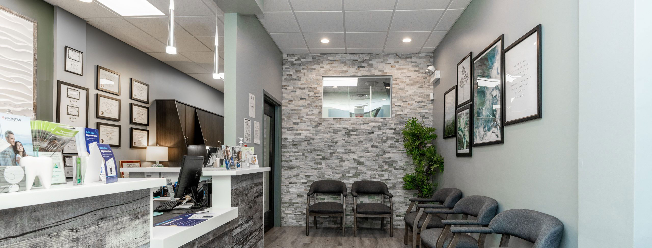
At Artistic Family Dental, we use digital radiography as a standard diagnostic tool because it supports thoughtful, evidence-based care while improving the overall patient experience. The technology complements routine exams and advanced procedures alike, providing a clear visual record that can be revisited whenever needed.
One of the biggest advances with digital imaging is the ability to view high-resolution images instantly. Instead of waiting for film to develop, the dentist and patient can review radiographs together during the same visit. This immediacy fosters clearer conversations about findings and treatment options, because the images are available for annotation and explanation on the spot.
Digital files can be enhanced without altering the original data, allowing practitioners to fine-tune contrast, magnification, and edge definition to reveal subtle changes. These enhancements improve diagnostic confidence for common concerns like interproximal decay, root morphology, and early periodontal bone loss. The result is better-informed clinical decisions and a more predictable treatment pathway.
Because images can be copied and stored easily, comparisons between past and present studies are straightforward. Tracking progression or healing over time becomes a practical part of preventive care and long-term maintenance, helping clinicians intervene earlier when trends suggest a problem.
Digital radiography generally requires less radiation than traditional film X-rays because of more sensitive sensors and more efficient image capture. Lower exposure contributes to patient safety while still delivering diagnostic-quality images. For those who require frequent monitoring—such as children or patients undergoing restorative or orthodontic treatment—this reduction can be especially meaningful.
Radiation safety remains a priority in every dental setting. Digital systems, combined with appropriate exposure settings and modern equipment, let clinicians tailor the dose to the clinical need. Protective measures such as lead aprons and thyroid collars are also used when appropriate to further minimize risk and provide patients with peace of mind.
Alongside dose reduction, digital radiography removes the need for chemical processing, which eliminates associated environmental hazards. The absence of developer fluids and paper-based workflows helps dental practices operate more sustainably while maintaining high standards of patient care.
Instead of film, digital radiography uses small electronic sensors placed in or near the mouth to capture X-ray images. There are several sensor types—such as intraoral sensors for bitewing and periapical images and extraoral detectors for panoramic studies—but all work on the same principle: convert X-ray energy into a digital signal. That signal is then processed and rendered as an image on a computer screen.
Once captured, images are stored securely in the patient’s electronic record. Digital storage enables efficient organization, rapid retrieval, and long-term archiving without the physical constraints of film. Files can be labeled, dated, and compared with prior studies to give a complete timeline of oral health, which is valuable for preventive care and restorative planning.
Sharing images with specialists or outside providers is straightforward because digital files are easily exported in standard formats. This streamlined transfer supports coordinated treatment—whether referring for endodontic evaluation, orthodontic planning, or surgical consultation—helping ensure every patient receives care informed by clear visual information.
For most patients, having a digital X-ray is a quick and comfortable experience. The dental team will position a small sensor in the mouth or align an external detector, then step briefly out of the immediate area to safely activate the X-ray. The whole process typically takes only a few moments, and the resulting image appears on the office monitor almost instantly for review.
Clinicians will often use those images to explain diagnoses and proposed treatments in real time, pointing out areas of concern and showing how proposed care will address them. This visual approach helps patients better understand their oral health and the rationale behind recommended procedures, fostering informed consent and shared decision-making.
If you have concerns about radiation exposure or the imaging process, bring them up with your dentist. The team can explain the safeguards in place, how digital radiography reduces exposure compared with older methods, and why certain images are necessary for comprehensive care. Clear communication helps patients feel comfortable and engaged in their treatment journey.
Digital radiography has become a cornerstone of modern dental care—faster, safer, and more adaptable than traditional film-based imaging. By combining instant visual feedback with secure digital records and simplified specialist collaboration, it supports accurate diagnoses and more efficient treatment planning. If you’d like to learn more about how digital X-rays are used in our office and what to expect at your next visit, please contact us for more information.


Digital radiography captures dental X-ray images using electronic sensors and computer processing instead of photographic film. The sensor converts X-ray energy into a digital signal that is rendered as an image on a monitor, which eliminates the need for chemical development and physical film storage. This modern workflow allows images to be viewed, adjusted, and stored immediately after capture.
Because images are digital from the start, clinicians can magnify, change contrast, and compare studies side by side without altering the original file. The process is typically faster and more efficient for both clinical teams and patients, enabling same-visit review and more streamlined record keeping. Digital capture also enables easy sharing with specialists and integration into electronic health records.
Digital images provide high-resolution detail that can be enhanced with software tools to reveal subtle changes in tooth structure and bone. Clinicians commonly adjust brightness, contrast and magnification to clarify areas of concern such as interproximal decay, root morphology and early periodontal bone loss. These capabilities increase diagnostic confidence and help clinicians identify problems that might be harder to see on film.
Instant access to prior studies also supports more accurate treatment planning by allowing direct comparisons over time. Being able to review images with patients during the appointment improves understanding of findings and fosters informed decision-making. Overall, digital radiography contributes to more predictable and evidence-based care pathways.
Digital radiography generally requires less radiation than traditional film because modern sensors are more sensitive and image processing compensates for lower doses. Clinicians select exposure settings appropriate to the patient's size and the diagnostic need, which helps minimize exposure while maintaining image quality. Protective measures such as lead aprons and thyroid collars are used when appropriate to further reduce risk.
Radiation safety is always a priority in dental imaging and digital systems are one component of an overall strategy that includes operator training and equipment maintenance. In addition to dose reduction, digital workflows eliminate chemical processing, reducing environmental hazards associated with developer solutions. Patients who have questions about exposure should discuss specific concerns with their dental team to understand the safeguards in place.
Most digital X-ray visits are quick and comfortable. A small intraoral sensor or external detector will be positioned in or near the mouth, the clinician will step aside briefly to activate the X-ray, and the image will appear on the office monitor within seconds. The entire exposure typically lasts only a moment and most patients experience little to no discomfort beyond sensor placement.
Once the image is captured, the dentist will review it on-screen and often use it to explain findings and recommended care in real time. This visual approach helps patients see the issue and understand treatment options, and it allows the team to answer questions immediately. If you have concerns about the process, mention them when you check in so staff can explain steps and accommodations.
After capture, digital radiographs are stored in the patient's electronic record where they are labeled and indexed for easy retrieval. Modern practice management and imaging systems organize files by date and type, making it straightforward to compare current images with prior studies and to track changes over time. Long-term archiving is handled electronically, removing the need for physical storage space used by film.
Image files are typically protected by access controls, user authentication and regular backups to preserve data integrity and availability. Secure transfer methods are used when sharing images with specialists or other providers to protect patient information. Patients who want to know more about record security can ask their dental team how imaging files are managed and safeguarded.
Yes, in many cases digital radiography can reveal early signs of disease sooner than film because of enhanced image quality and post-capture manipulation. Adjusting contrast, magnification and edge enhancement can make small lesions, initial interproximal decay and subtle bone changes more apparent. Early detection supports less invasive treatment and better long-term outcomes.
Frequent or periodic digital imaging also makes it easier to monitor trends and healing over time, which is particularly useful for preventive care and restorative follow-up. When clinicians see subtle progression, they can intervene earlier to slow or stop disease. That longitudinal perspective is an important advantage of a digital imaging workflow.
Digital intraoral and extraoral radiographs provide excellent two-dimensional information but have limits when a three-dimensional view is required. For complex implant planning, certain surgical assessments or detailed evaluation of root anatomy, clinicians may recommend cone beam computed tomography (CBCT) or other advanced imaging that offers volumetric detail. Likewise, overlapping structures or patient positioning can obscure findings on plain radiographs.
Artifacts from metal restorations or patient movement can also affect image quality and may require repeat exposures or alternative techniques. Clinicians weigh the diagnostic benefit against exposure and will order additional imaging only when it adds clear value to diagnosis or treatment planning. Clear communication with the dental team helps patients understand why a specific study is recommended.
Digital files can be exported in standard formats that specialists can review on their own systems, which streamlines referrals and collaborative case planning. Sending images electronically reduces the need to transport physical film and allows receiving clinicians to evaluate studies quickly and provide timely input. This efficiency helps maintain continuity of care for procedures such as endodontics, oral surgery and orthodontics.
Many practices use secure portals or encrypted file transfer to share radiographs while protecting patient privacy. Rapid access to high-quality images enables more precise consultations and can shorten the time between diagnosis and definitive treatment. Patients benefit from coordinated care that is informed by clear visual information.
Frequency of dental X-rays is not the same for every patient and is determined by individual risk factors, clinical findings and treatment needs. A dentist will consider your oral health history, the presence of symptoms, age, restorative work and periodontal status before recommending an imaging schedule. For routine preventive care, imaging intervals are tailored to balance diagnostic benefit with the principle of using the lowest reasonable exposure.
Children, patients with active disease or those undergoing certain treatments may require more frequent monitoring, while low-risk adults with stable oral health may be imaged less often. Discussing your specific circumstances with the dental team ensures the imaging plan supports safe, effective care that is appropriate for your needs.
Artistic Family Dental uses digital radiography to combine high-quality diagnostic imaging with a patient-centered experience that emphasizes speed, clarity and safety. Digital images allow clinicians to make informed decisions more quickly, to show patients exactly what they see, and to maintain detailed records that support long-term monitoring. The workflow also reduces environmental impact by eliminating chemical processing and physical film handling.
By integrating digital radiography into routine exams and complex procedures, the practice enhances diagnostic precision and facilitates collaboration with specialists when needed. Patients benefit from faster appointments, clearer explanations of findings and the ability to track oral health over time through easily accessible digital records.
Ready to schedule your next appointment or learn more about our services?
Our friendly team is here to make it easy. Whether you’d like to call, email, or use our convenient online form, we’ll help you find the right time and answer any questions you have. Don’t wait to take the next step toward a healthier, more confident smile—contact Artistic Family Dental today and experience the difference genuine, personalized care can make.

Back to top